Generic selectors
Exact matches only
Search in title
Search in content
Post Type Selectors

Departments: Tenure-Track Faculty

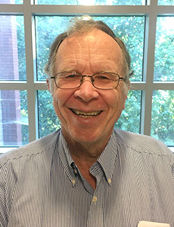
Michael G Hahn

Short Biography:
Dr. Hahn received a B.S. in chemistry and a B.A. in Independent Studies in 1974 from the University of Oregon and his Ph.D. in biochemistry in 1981 from the University of Colorado. A postdoctoral research associate appointment at the University of Wisconsin-Madison in plant pathology followed, after which Dr. Hahn went to the Albert-Ludwigs-Universiät (Freiburg, Germany) with the support of an Alexander-von-Humboldt stipend. Following another postdoctoral research associate appointment at the Salk Institute (San Diego, CA), Dr. Hahn joined the CCRC in July 1986. Full publications: 77.

Research Interests:
Our laboratory studies the cell biology and biosynthesis of plant cell walls. Plant cell walls play major roles in the biology of plants. Examples of these roles include controlling the growth and shape of plant cells, tissues, organs, and ultimately the entire plant, regulating the movement of nutrients and signals within the apoplast and toward the plasma membrane, serving as the first line of defense against pathogens and environmental stresses, and acting as a source of signaling molecules important in plant development and defense. Plant cell walls are also the principal component of plant biomass, which has become a focal point in the search for alternative and renewable sources of energy (biofuels).
We are pursuing two broad research goals:

(A) We are investigating plant cell wall biosynthesis by looking at two families of genes, primarily in Arabidopsis, thought to encode glycosyltransferases involved in plant cell wall glycan biosynthesis: 1) GAlacturonosylTransferase-Like (GATL) proteins thought to be involved in pectin biosynthesis; 2) FUcosylTransferase (FUT) proteins thought to add fucosyl residues to diverse plant cell wall glycans.

(B) We have developed a large and diverse library of monoclonal antibodies against plant cell wall glycans. These antibodies are being used to determine the locations of diverse cell wall carbohydrate structures (epitopes) in Arabidopsis, switchgrass and poplar. These antibodies are also proving useful for plant cell wall mutant characterization studies, and for quantitating glycans in cell wall extracts.

Our laboratory utilizes a broad range of experimental approaches in these studies, including molecular genetic, biochemical, immunological and microscopic techniques.

Google Scholar

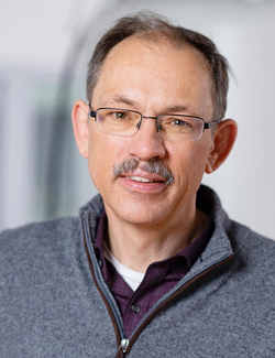
Art Edison

Short Biography:
Art Edison is a Georgia Research Alliance Eminent Scholar and Professor of Genetics and Biochemistry and a member of the Complex Carbohydrate Research Center and Institute of Bioinformatics at the University of Georgia. He received his Ph.D. in biophysics from the University of Wisconsin-Madison, where he developed and applied NMR experimental and theoretical methods for protein structural studies under the supervision of John Markley and Frank Weinhold. He joined the faculty at the University of Florida and the National High Magnetic Field Laboratory in 1996. He advanced from Assistant to Full Professor in the UF Department of Biochemistry & Molecular Biology. Prof. Edison was the founding PI and Director of the NIH-funded Southeast Center for Integrated Metabolomics, and his research focuses on the role of small molecules in biology and disease. In 2015, Edison moved to the University of Georgia where he directs the CCRC NMR facility, which supports research in both metabolomics and structural biology. Edison’s research group collaborates on several metabolomics projects from microbes to humans.

Research Interests:
The Edison lab uses metabolomics to solve problems in biology and biomedicine. Metabolomics is the omics technology that focuses on metabolites in the context of systems biology. As such, we spend a considerable amount of effort on bioinformatics, data integration, and modeling of data. Our primary analytical technology is NMR, but we also regularly use mass spectrometry. We develop new NMR and bioinformatic methods and collaborate extensively with other groups that have interesting applications or complementary technologies.

Google Scholar

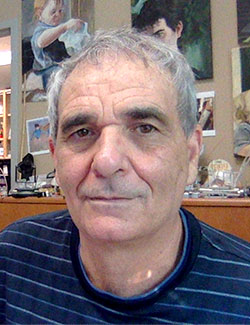
Alan Darvill

Short Biography:
Alan Darvill received his B.S. in plant biology in 1973 from Wolverhampton Polytechnic (England) and his Ph.D. in plant physiology in 1976 from the Aberystwyth (Wales). He founded the CCRC with Dr. Peter Albersheim in September 1985. Dr. Darvill is Director Emeritus of the CCRC, former Director of the Department of Energy (DOE)-funded Center for Plant and Microbial Complex Carbohydrates, and was University of Georgia lead in the DOE-funded BioEnergy Science Center. He was elected chairman for 1994-95 of the Carbohydrate Division of the American Chemical Society, and was appointed a member in 1993 and chairman in 1996 of the Martin Gibbs Medal Committee of the American Society of Plant Physiologists. He served on the editorial boards of Glycobiology and of the Plant Journal for Cell and Molecular Biology. Dr. Darvill received the Outstanding Faculty Award of the UGA Chapter of the Golden Key National Honor Society in 1995. In 2003, Dr. Darvill was appointed Regents Professor of Biochemistry and Molecular Biology and Senior Faculty Fellow. In 2008, he was appointed to the University of Georgia Research Foundation Board, and in 2010 was named Fellow of the American Association for the Advancement of Science. Full publications: 220.

Research Interests:
Characterization of the non-cellulosic polysaccharides that constitute the primary cell walls of plants has been a major research goal of our laboratory for many years. We have emphasized the structural studies of five major non-cellulosic polysaccharides, namely, the pectic polysaccharides rhamnogalacturonan I (RG-I), rhamnogalacturonan II (RG-II), and homogalacturonan, and the hemicellulosic polysaccharides xyloglucan and arabinoxylan. We have characterized the structures of these polysaccharides isolated from angiosperm and gymnosperm cell walls. We have come to realize that these polysaccharides are the predominant non-cellulosic polysaccharides of cell walls. These polysaccharides, although varying in quantity in the cell walls of angiosperms and gymnosperms, are structurally conserved, indicating their importance in primary cell wall structure and function.

These cell wall, non-cellulosic polysaccharides represent a group of carbohydrates with considerable structural complexity that offer a major challenge to those wishing to decipher their structures. RG-II, for example, is probably the most structurally complex polysaccharide so far isolated from nature. RG-II was first discovered to be a component of the primary cell walls of plants by our laboratory in 1978 and has now been found in the walls of angiosperms, gymnosperms, lycophytes, and pteridophytes. RG-II is a small polysaccharide consisting of approximately 30 glycosyl residues. However, this polysaccharide contains at least 12 different glycosyl residues linked together in over 20 different glycosyl linkages. RG-II is characterized by several unusual sugar components including 2-O-methyl fucose, 2-O-methyl xylose, apiose, 3-C-carboxy 5-deoxy-L-xylose (aceric acid), 3-deoxy-D-manno-2-octulosonic acid (KDO), and 3-deoxy-D-lyxo-2-heptulosaric acid (DHA). Both aceric acid and DHA have been found in no natural source other than RG-II.

We have characterized oligosaccharide fragments of RG-II that contain 28 of the possible 30 glycosyl residues of this pectic polysaccharide. We have obtained evidence that the backbone of RG-II consists of seven a-1,4-linked galactosyluronic acid residues to which two disaccharide, one heptasaccharide, and one octasaccharide side chains are attached. These side chains are attached to the backbone via the acid labile ketosidic and glycosidic linkages of KDO, DHA, and apiose. The demonstration that RG-II exists in primary walls as a dimer that is covalently cross-linked by a borate diester was a major advance in our studies of this polysaccharide. Dimer formation results in the cross-linking of the two homogalacturonan chains upon which the RG-II molecules are constructed and is required for the formation of a three dimensional pectic network “in muro” (Annual Review of Plant Biology, 2004, 55:109-139).

Research in our laboratory continues to emphasize the structural characterization of the non-cellulosic polysaccharides of cell walls. The availability of cell wall mutants is assisting us in these structural studies. Structural analysis is accomplished by release of polysaccharides from cell walls and generation of defined polysaccharide fragments with purified enzymes or chemical treatments, chromatographic purification of the oligo- and polysaccharides, chemical structural characterization techniques, and a combination of several analytical techniques including gas-liquid chromatography, liquid chromatography, gas-liquid chromatography-mass spectrometry, liquid chromatography-mass spectrometry, MALDI-TOF mass spectrometry, and 1H- and 13C-NMR spectroscopy.

We are also collaborating with Dr. Michael Hahn in the production of monoclonal antibodies to cell wall polysaccharides and the oligosaccharides derived from these polysaccharides. These antibodies will be used in both purification of oligosaccharides and, more importantly, to begin to decipher the arrangement and location of the polysaccharides within the primary cell wall using monoclonal labeling of specific epitopes on the wall polysaccharides.

Russell W Carlson

Short Biography:
Dr. Carlson received his B.A. in chemistry from North Park College, Chicago, Illinois in 1968, and his M.S. and Ph.D degrees in biochemistry from the University of Colorado in 1974 and 1976, respectively. Dr. Carlson is Emeritus Professor of Biochemistry and Molecular Biology (BCMB), and of the Complex Carbohydrate Research Center (CCRC) at the University of Georgia (UGA), Athens, Georgia. Prior to retiring in 2014, Carlson was Professor of BCMB, Executive Technical Director of Analytical Services at the CCRC, and Adjunct Professor of Microbiology at UGA. His area of research is microbe-host interactions; specifically, the role that microbial cell surface carbohydrates have in determining the virulence of both animal and plant pathogens (and symbionts). He has nearly 200 peer-reviewed publications and his research was funded over the years by grants from the National Institutes of Health, the National Science Foundation, the United States Department of Agriculture, as well as contracts with the Centers for Disease Control.

Research Interests:
Research in Dr. Carlson’s group is focused on characterizing the molecular basis for the interaction between a bacterium and a plant or animal host cell. One system being studied is the symbiotic, nitrogen-fixing infection of host legumes by rhizobia bacteria. Carlson’s group also has projects directed toward characterizing the roles bacterial lipopolysaccharides (LPSs), lipooligosaccharides (LOSs) and capsular polysaccharides (CPSs) play in determining pathogenicity (in animal hosts) of such organisms as Salmonella enteritidis, Neisseria meningitidis, Hemophilus influenzae, Moraxella catahalis, and, recently, Bacillus anthracis. Both the plant symbiont and animal pathogen work are being undertaken in collaboration with several research groups in other universities. The following paragraphs briefly describe several of these projects.
The nitrogen-fixing symbiosis is a complex infection process in which the soil bacteria contain genes that are activated by flavonoid molecules produced by the host plant. These genes encode enzymes that synthesize a glycolipid, which is an acylated chitin oligosaccharide. In most cases, the glycolipids produced by each Rhizobium species are structurally modified, which results in their ability to interact only with a specific legume plant (e.g., Rhizobium leguminosarum biovar viciae infects peas but not alfalfa, while R. meliloti infects alfalfa but not peas). This molecular “recognition” process results in the stimulation of cell division in the legume root causing a nodule to form. The cells in this nodule are invaded by the rhizobia and are where nitrogenase is produced that reduces dinitrogen to ammonia. Other molecules on the surface of rhizobia are required for invasion of the root nodule cells by these bacteria. These are the outer membrane LPSs and CPSs. Specific structural changes occur in these molecules in response to the host plant which are crucial for infection. These structural changes and the genes responsible for them are presently under investigation by Carlson’s research group. Knowledge gained in understanding the molecular basis for Rhizobium-legume symbiosis may lead to improving the yield of important legume crops and increasing the fertility of soil for non-legume crops. Besides the potential benefit to crop yield and soil fertility that may be gained from new knowledge about Rhizobium-legume symbiosis, these studies also have a bearing on how the plant’s defense mechanism is regulated so that the growth of the bacteria is controlled by the host to establish a symbiotic rather than a pathogenic relationship. Details of this work are described in several relevant publications that are referenced below.

Carlson’s group, in collaboration with Dr. David Stephens at Emory University, has also worked on determining the structural basis for the virulence functions of the cell surface lipooligosaccharide (LOS) and capsular polysaccharide (CPS) from Neisseria meningitidis. These molecules contain novel structural features that inhibit the host’s defense response. In the case of the LOS these structural features include the synthesis of host-like structures to camouflage the bacterium, as well as modifications that inhibit serum killing, etc. The CPS structures also protect the bacterium from the host defense reponse as they are poorly immunogenic. However, it is known that isolated CPS can be conjugated to proteins and have proven to be effective vaccine antigens that prevent infection by several types of N. meningitidis. Thus, the objective of this work is to identify the virulence functions of LPS and CPS structures as well as identify optimal structures for the development of vaccine antigens. Several publications describing this work are in referenced below.

Carlson’s group, in collaboration with Dr. Conrad Quinn at the Centers for Disease Control and Dr. Geert-Jan Boons of the Complex Carbohydrate Research Center at UGA, is working on determining if there are potential carbohydrate structures in the cell wall of Bacillus anthracis that can be used for the development of therapeutics, vaccines, and diagnostics. Ever since the anthrax attacks surrounding the events of 9/11, there has been, as part of the biodefense initiative, a great deal of focus on the development of novel therapeutics, vaccine antigens, and diagnostic agents for the treatment, prevention, and identification of B. anthracis infections. The cell surface polysaccharides that surround the bacterial cell or are part of its cell wall are well documented virulence factors, they have been used for the preparation of commercially available vaccines for the prevention of a number of bacterial diseases, and they are also known to be the basis for the serotyping of numerous bacterial pathogens; both Gram-positive and Gram-negative. Therefore, B. anthracis cell wall carbohydrates were investigated to determine if they also have potential in these areas. Bacillus anthracis is a member of the B. cereus group of bacteria; all of which are quite closely related as determined by comparing genome sequences. Laboratory-grown cultures of B. anthracis produce very few cell wall carbohydrates. In fact, only two major carbohydrates have been characterized. One is from a coating that surrounds the spore, called the exosporium. It is an oligosaccharide that covers a collagen-like protein called BclA. The structure of this oligosaccharide was determined by the group of Charles Turnbough at the University of Alabama, Birmingham. This structure, various structural analogs, and their protein conjugates have been synthesized by the group of Geert-Jan Boons at the CCRC and immunochemically characterized in collaboration with Conrad Quinn at the Centers for Disease Control. A second is a cell wall polysaccharide from the vegetative cell wall of B. anthracis. The structure of this polysaccharide as well as that from closely related B. cereus strains that are pathogenic have been characterized at the Complex Carbohydrate Research Center. Both the structural and immunochemical analyses of these cell wall carbohydrates support their potential for the development of vaccines and diagnostics. In addition, the data also indicate that these carbohydrates may be important for the virulence of this pathogen.

Dr. Carlson’s research has been supported by the U. S. Department of Agriculture, the National Science Foundation, and the National Institutes of Health.
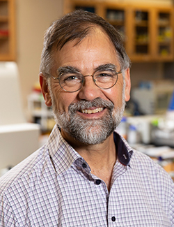
Geert-Jan Boons

Short Biography:
Dr. Boons received his B.S. in Chemistry in 1983 and his Ph.D. in Synthetic Carbohydrate Chemistry in 1991 from the State University of Leiden (Netherlands). Prior to joining the faculty at the CCRC in 1998, he spent seven years in the United Kingdom, first as a postdoctoral fellow at Imperial College, London, and the University of Cambridge, and then as a lecturer and professor at the University of Birmingham. In 2003, Dr. Boons was awarded the Carbohydrate Research Award for Creativity in Carbohydrate Science by the European Carbohydrate Association. Also in 2003, he was elected chairman for the 2005 Gordon Research Conference on Carbohydrates. He serves on the editorial boards of Carbohydrate Research , the Journal of Carbohydrate Chemistry , and Advances in Carbohydrate Chemistry and Biochemistry. In 2004, Dr. Boons received the Horace Isbell Award by the Division of Carbohydrate Chemistry of the American Chemical Society and was appointed Franklin Professor of Chemistry in the College of Arts and Sciences at the University of Georgia.

Research Interests:
The research of the Boons Group deals with the synthesis and biological functions of carbohydrates and glycocongugates. The diversity of topics to which the group has significantly contributed include the development of new and better methods for synthesizing exceptionally complex molecules, the use of new methods in the synthesis and study of properties of complex carbohydrates of increasing size and complexity, the development of synthetic cancer and bacterial vaccines, the design and synthesis of glycosidase inhibitors and the use of synthetic compounds for the study of innate immunity

Maor Bar-Peled

Short Biography:
Dr. Bar-Peled received his B.S. in 1985 and his M.S. in 1988 from the Hebrew University of Jerusalem. He completed his Ph.D. studies in 1993 in the Department of Plant Genetics, Weizmann Institute of Science in Israel. In 1992, Dr. Bar-Peled was a recipient of the Science Prize given by the Feinberg Graduate School of the Weizmann Institute of Science and, in 1993, he received an Israeli Ministry of Education Award. Prior to coming to the University of Georgia, Dr. Bar-Peled spent five years in the Department of Energy Plant Research Laboratory, Michigan State University, as a postdoctoral fellow. Dr. Bar-Peled was also a visiting scientist in the Department of Molecular Biology at the Washington University School of Medicine in St. Louis.Full publications: 32

Research Interests:
Research in Dr. Bar-Peled’s group aims to understand, at the molecular level, the roles of complex glycans in living organisms. We are interested in the roles of cell surface glycans in cell-cell recognition, pathogenicity, and communication between micro-organisms and their plant or animal hosts. In addition, we are investigating how the cellular processes involved in the synthesis, regulation and assembly of plant cell walls can be modified to enable new cost-effective technologies for producing biofuels from plant biomass. Our research uses biochemical, molecular and cellular and bioinformatics techniques together with plant and microbial mutants and state of the art mass spectrometry and NMR spectroscopy.

Current research programs in the Bar-Peled lab are:

The role of cell surface glycans during the life-cycle of Rhizobium. We study the molecular events that trigger this free-living soil bacterium to alter its cell surface glycan and glycolipid composition in response to changes in its environmental.

The molecular mechanisms that allow Bacillus cereus to form spores that adhere to diverse surfaces. This common soil bacterium is a difficult to control food poisoning agent.

The relationship between cell surface glycan synthesis and the interactions between fungi and their plant and animal hosts. We study how fungi adhere to and penetrate host cells and how this is related to diseases caused by fungi.

The genes and enzymes involved in the synthesis of plant cell wall glycans. We study how glycan synthesis is regulated and how these glycans are formed in the Golgi and then transported to plasma membrane where they are assembled into a functional wall. Understanding such processes at a molecular level will enable the development of bioenergy crops that can be cost-effectively converted to liquid fuels.